Vital Pulpotomy In A dog
Above: The root canal is being filled with a flowable veterinary dental composite. The pet is under anesthetic at the veterinary dental clinic.
Above: The final repair

At home with a repaired canine tooth
Clinical signs of veterinary dental tooth fractures
- Usually no clinical signs they are subtle and painful initially
- Bleeding from tooth in the first 24 hours
- Molar abscess below the eye especially associated with a fracture of the maxillary premolar 4 or carnassial tooth ( a large three rooted tooth
- A dental explorer – pointed to help determine pulp exposure- pulp exposure after a tooth fracture usually means pain, and needing a vital pulpotomy or root canal or extraction of the affected tooth.

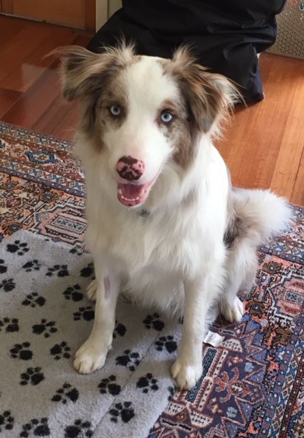
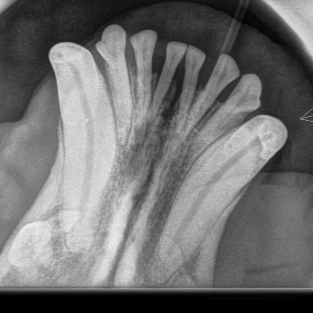
Bowie a 6 year old Staffie Cross fractured a canine tooth and was seen by the veterinary dentist straight away, A vital pulp capping was done at the vet dental clinic. This is an X-ray of the bottom jaw teeth, the canines on the outside and incisors on the inside. The arrow is over the repaired area. This was done in October 2018.
Assessment
Our first veterinary dental exam is done in the consult room on an awake dog , and an initial assessment is done with the recommended therapy.
Many of the dental procedures we do are second opinions dentals with the dental procedure done on the first visit, on the same day. Owners book in a consultation – we review the dental disease – and with informed consent do a dental procedure under general anesthesia on the same day.
Veterinary dental exam to determine outcome
Vet Dental radiology is becoming more common – but adds to the costs –
Pathology of the veterinary dental tooth roots – peri apical disease, and vet dental abscess are often not visible with out an X-ray
A proper vet dental exam, and dental x-rays can only be done under General anesthetic.
Anatomy of the tooth – Dental Veterinarian
- the crown of the tooth – the part in the mouth, above the gum line is surrounded by enamel
- the hard outer coating of the teeth that we see is Enamel – it is not a sensitive tissue – it is the hardest tissue in the body and once damaged exposes dentin that is rough and they will pick up plaque, and is sensitive – damaged enamel does not repair itself –
- under the enamel is the Dentine or Ivory – and can regenerate to some degree
- then the pulp chamber or nerve of the tooth – the living tissue of the tooth.
Classification of tooth fractures
enamel infraction – from chewing – superficial -treatments seal with resin
enamel fracture- good prognosis – loss of enamel without dentine exposed – treat with resin
uncomplicated crown fracture – pulp not exposed = seal off with resin – may restore – good prognosis
Complicated crown fracture -if new vital pulpotomy OR root canal Or extraction = only three options. DO NOT LEAVE
etc
We often extract a tooth rather than choose to do repeated anesthetics to save a tooth.
Root canal- vital pulpotomy procedure – dog dental
The pulp contains nerves and blood vessels and can bleed and be painful when damaged. A vital pulpotomy is one of the few emergency treatments that are performed in veterinary dentistry. It provides the only treatment option, besides extraction, or root canal for freshly exposed pulp . This treatment, by Vet dentists, aims to preserve the vitality of the pulp of the affected tooth, allowing it to continue its growth and remain healthy
Veterinary dentists try to wait longer ( 9 -12 months), before doing an elective vet dental pulpotomy, Dog dentists wait as long as you can (for tooth maturation) but not too long (before one gets permanent damage to the palate and/or upper canines). Therefore, recent injuries in teeth of relatively young dogs are the best candidates for vital pulpotomy by a Dog dentist.
Keep the area sterile – remove pulp – pulp dressing – placement of the restoration
- Isolate clean with a latex glove , dam and sterilize the area -This surgical procedure requires sterile technique.
- Reshape the damaged tooth – usually involves a crown amputation with a vet dentist – tapered fissure bur.
- Amputate pulp – brand new high speed round burr, about 4 -6 mm depth
- Should be bleeding from pulp as it is vital 0-> Gently wash the pulp with saline
- Dog dentists use paper points – swabs for teeth -placed into opening -> to control bleeding takes about 5 minutes – also sterile saline flush.
- Calcium hydroxide is applied by the dog dentist – 1 -2 mm thick pulp dressing – onto the exposed pulp, encouraging the formation of a tertiary dentine bridge.r
- Base “filling” with a glass ionomer – 1 mm layer is placed over the CA2(OH)3 dressing
- Final restoration -by dental veterinarian is -with a routine composite restoration material ( phosphoric acid etch first – bonding interface layer -composite material- file smooth)
- Post op x ray 4 months later – a dental bridge formed if all going well.
